

NEW
Mobile Ultrasound
Home visits now possible.
Languages:
German, English, Farsi, French
Appointments:
Tel.: +43 670 3529636
Email: contact@ultraschallwien.at
Dr. Negar Fakhrai
Specialist for radiology
Languages:
German, English, Farsi, French
Appointments:
Tel.: +43 670 3529636
Email: contact@ultraschallwien.at
Home visits possible.

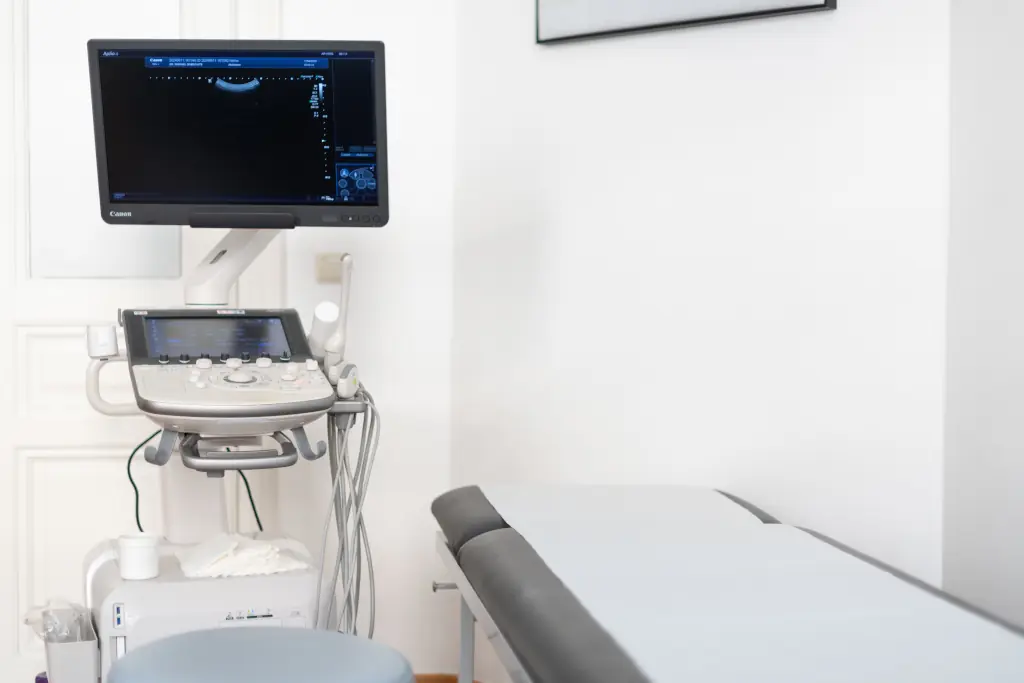
Ultrasound can be used frequently and in many different ways.
This examination method is primarily non-invasive, does not require X-rays and therefore does not involve any radiation exposure.
Furthermore, an ultrasound examination is usually painless, without side effects and in any case harmless. For this reason, this method is also ideal for all age groups and pregnant patients.
CONTACT DETAILS
UROLOGIE DR. DOBROVITS
Martinstraße 91/8a
1180 Vienna
Dr. Fakhrai - Contact details:
Tel.: +43 670 3529636
Email: contact@ultraschallwien.at
Google Maps


SERVICES
Ultrasound examination in:
Preventive medicine
- Skin and subcutaneous tissue
- Carotis-Duplex-Sonographie
- Vein-Duplex-Sonography
- Abdominal Aorta
- Thyroid gland, salivary glands, head and neck lymphnodes
Aftercare
- Upper & lower abdomen
- Vessels
- Lymph node stations | for diseases such as melanoma, lymphoma or breast carcinoma
Follow-up
- Progression, Regression & improvement of various findings and clinical pictures (i.e. spleen size in EBV infection, lymph node size and morphology, post-traumatic follow up of haematomas)
Initial diagnosis / to clarify complaints within the:
- Abdominal wall and groin
- Muscles and joints (shoulder, elbow, wrist, hip, knee, ankle, palm, sole of the foot, etc.)
- Abdomen (upper abdomen, kidneys/retroperitoneum, lower abdomen)
Urology
- Kidneys, Kidney transplant (NTX)
- Urinary bladder and residual urine
- Testicles
Gynaecology
- Uterus & Ovaries
- Breast including armpits on both sides in women & men
Paediatrics
Ultrasound is an indispensable radiological examination method in paediatrics.
- Abdomen / Appendix
- Hip
- Lymphnodes
Operation release
Depending on which operation is planned, different examinations are necessary in the course of preparation, often including ultrasound examinations.INFORMATION
Breast ultrasound incl. armpits on both sides
It is important to mention that an ultrasound examination alone is not suitable for the early detection of breast cancer.
 From the age of 40, women in Austria are invited to have a mammogram and, depending on the density of the mammary gland, a supplementary ultrasound scan of the breast on both sides every other year as part of the early detection of breast cancer (BKF).
Breast ultrasound can be carried out at any age, even if there are questionable changes at a young age. It can also be used regularly and often without hesitation, for example to follow up the progression of benign changes.
Men should also have any changes in their breasts such as swelling, lumps, increase in size, etc. followed up.
F.A.Q.
Frequently asked questions concerning ultrasound examination
Basics
An ultrasound examination, also known as sonography, uses sound waves to make the inside of the body visible. Ultrasound waves are sent into the body using an ultrasound device via a transducer and, depending on which organ these waves hit, they are reflected in different ways and received again by the transducer. An image is then calculated.
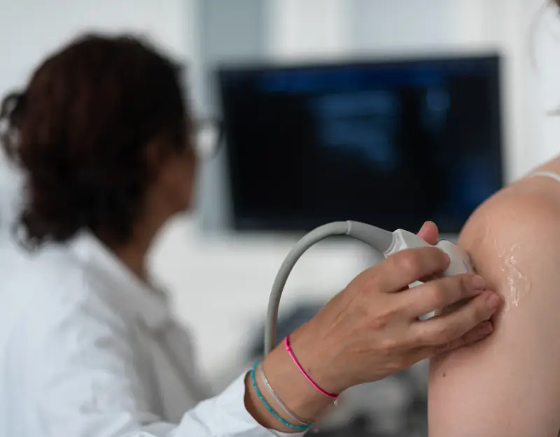 This method can be used frequently and in a variety of ways, is primarily a non-invasive examination method and does not require X-rays, so it does not involve any radiation exposure. Furthermore, an ultrasound examination is usually painless, without side effects and, in any case, harmless. For this reason, this method is also ideal for our very young patients. Gynecologists use ultrasound in pregnancy care so that any pathologies (diseases) of the baby can be detected intrauterine (in the uterus).
Which organs can be examined using ultrasound?
In principle, ultrasound can be used to examine all organs, even of an unborn baby. Exceptions are organs filled with air such as the lungs and the gastrointestinal tract as well as bones, as ultrasound waves ‘cannot pass through’ or are completely ‘reflected back’ by air and bone.
I am pregnant. Can I still have an ultrasound scan?
Yes, of course. There is no risk for the baby from the ultrasound. The direct examination of the unborn child in the uterus as carried out by gynecologist during pregnancy also has no harmful effect.
What do I need to consider before an ultrasound scan?
Upper abdominal ultrasound:
We ask you not to eat for at least 6 hours before the examination. This is to ensure that the gallbladder has not collapsed postprandially (emptied gallbladder after eating) and that there is not too much air in the stomach and intestines, which would make the examination conditions more difficult and restrictive. We ask you to take your medication with a sip of non-carbonated water. Please do not drink coffee or smoke a cigarette. Tea is possible, however it should be unsweetened. It is best to avoid flatulent foods the evening before.
Ultrasound of the lower abdomen:
We ask you to come with a full bladder (tap water or still water), as an empty bladder cannot be assessed.
Carotid duplex sonography / sonography of the neck organs:
For men, a fresh shave optimises the quality of the examination.
What kind of ultrasound examinations are there?
Ultrasound surface:
This refers to the skin and subcutaneous tissue. There are often changes such as ‘swelling’ or ‘lump formation’ in a certain area of the body. These can grow slowly over a long period of time if they are benign, for example. Post-traumatic after an accident of any kind, superficial changes can also occur suddenly. There are also fast-growing changes on the surface, for example in malignant diseases.
In our everyday work, we typically diagnose benign neoplasms (new formations) such as a lipoma (fatty tumour) or atheroma (sebaceous gland). In the case of post-traumatic swelling, ultrasound often reveals fluid accumulation in the form of haematomas (bruising) or diffuse oedema (extensive swelling/fluid retention). Malignant diseases can lead to skin or lymph node metastases from the primary tumour (metastases from the original malignant cells).
Ultrasound lymph node sites:
We humans have around 600 lymph nodes which, together with the spleen and tonsils, belong to the secondary lymphatic organs and are distributed throughout the body as a lymphatic system. This system plays an important role in our immune system.
The lymph nodes are distributed quite superficially, for example in the head/neck area - 50% of our lymph nodes are localised here. In the area above and below the collarbone on both sides, in the armpits on both sides and also in the groin on both sides. These superficial lymph nodes are easy to visualise with ultrasound.
Their number, size and shape are examined and can often be used to differentiate between normal, inflammatory or malignant lymph nodes. Ultrasound examination of the lymph node sites is important in the planning of operations and aftercare for melanoma (skin cancer). It is also important in the planning of operations for malignant diseases in the head and neck area, for example in the case of thyroid carcinoma.
This examination is also common in the clarification of haematological diseases (e.g. of the haematopoietic bone marrow or the lymphatic system).
Ultrasound of the large neck vessels supplying the brain/carotid duplex sonography:
For this examination, ultrasound/sonography and Doppler sonography are used, which together produce a cross-sectional image of the vessel as well as visualising and measuring the blood flow in the vessel. The arteria carotis communis, the arteria carotis interna, the arteria carotis externa and the arteria vertebralis can be examined on the left and right.
Various diseases can alter the arterial vessels (which supply the body's cells with oxygen and nutrients, among other things) and subsequently lead to stenoses (constrictions) due to plaque deposits and calcifications which, the more they progress, can have dangerous consequences for the patient, for example an increased risk of stroke.
Untreated high blood pressure, untreated high blood lipids, lipometabolic disorders, diabetes, smoking/nicotine abuse are some examples than can pathologically alter/constrict the arterial vessels.
 With Doppler ultrasound, it is possible to recognise whether there are pathological plaques (deposits) in the vessels and to what extent the lumen (diameter) of the vessel is impaired/ narrowed.
During this examination, the intima-media thickness (thickness of the inner lining of the vessel) is measured, which is an important indicator of fat and calcium deposits in other arteries (for example in the heart - coronary heart disease [CHD] or in the legs - intermittent claudication [PAOD]).
This examination is necessary in the course of various surgical preparations/operation authorisations.
Furthermore, aneurysms (pathological dilation of the arteries) can be diagnosed, which in some cases need to be followed up regularly and in some cases surgically repaired.
Ultrasound of the deep leg veins / venous duplex sonography:
This examination is primarily used to rule out or detect and assess the extent of deep vein thrombosis, or DVT for short (blood clots in the deep veins of the legs). It can also diagnose thrombophlebitis (inflammation and clots in the superficial veins) or venous insufficiency in varicosity (leaking venous valves due to varicose veins) or post-thrombotic syndrome (after thrombosis), which often leads to leg oedema (swollen legs/feet).
Ultrasound of the thyroid gland:
An ultrasound of the thyroid gland involves measuring the size of the organ, assessing the shape and the nature of the tissue. It plays an important role in the assessment of any cysts and nodules, which can be recognised from a size of just a few millimetres. There are numerous characteristics in ultrasound that enable changes in the thyroid tissue to be categorised and any further clarification that may be necessary (for example by means of blood analysis, nuclear medicine or biopsy/sampling) to be initiated. There are some clinical pictures that show typical images on ultrasound. For example, autoimmune thyroid disease - Hashimoto's disease. Ultrasound of the thyroid gland is used in follow up / post-operative follow up.
Ultrasound of the neck organs/ soft tissues of the neck:
In this examination, the parotid gland and submandibular gland (large parotid glands) are examined on both sides. Their size and condition are assessed. There are chronic inflammatory changes, some of which are caused by autoimmune diseases. Then there are concretions (salivary stones) that cause ductal obstruction and oedema (obstruction of an excretory duct, ductal dilatation, accumulation of saliva and swelling) which can be very painful. There are benign and malignant changes within these glands and the ultrasound features are used to recognise the need for further clarification.
Ultrasound abdominal wall and groin:
The most common hernia inguinalis (inguinal hernia) or hernia epigastrica (abdominal wall hernia/upper abdominal hernia) or hernia cicatricea (incisional hernia). This is a weakness and consecutively a gap in the abdominal wall/scar, whereby subcutaneous fatty tissue, peritoneum and intestines can protrude through the gap.
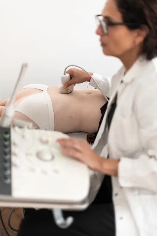
Ultrasound of the upper abdomen:
The liver, gallbladder, pancreas and spleen are located in the upper abdomen. In addition, lymph nodes and the large blood vessels, the abdominal aorta and inferior vena cava, can be seen. It is possible to recognise whether the organs are within the normal range in terms of size and structure or whether changes are present as part of various clinical pictures. For example, splenomegaly (enlargement of the spleen) in the case of an infection with Ebstein-Barr virus (EBV).
Ultrasound of the kidneys:
The kidneys are located retroperitoneally (behind the peritoneal cavity/behind the peritoneum). Whether the kidneys are in a normal position, of normal shape and size, whether there is hydronephrosis (urinary retention) or other abnormalities can be examined by ultrasound.
Ultrasound kidney transplant (NTX):
If the kidneys on both sides no longer function due to advanced kidney disease, a kidney transplant can be performed. The transplanted kidney is then regularly examined using blood analysis/renal function parameters and ultrasound.
Ultrasound residual urine:
In this examination, the volume of urine in the bladder is measured post-micturition (immediately after urination). This should always be less than 100ml.
Ultrasound lower abdomen:
The urinary bladder and lower abdominal organs (uterus, ovaries and prostate) are examined for abnormalities. Diseases of the appendix such as appendicitis (inflammation of the appendix) can also be diagnosed. If the inflamed appendix itself cannot be seen, the clinical symptoms (typical pain, fever, nausea and vomiting) and ultrasound features such as ascites/free fluid and/or lymphadenopathy can be used to draw conclusions and promptly initiate further surgical clarification.
Ultrasound muscles and joints (shoulder, elbow, wrist, hip, knee, ankle, palm, sole of the foot, Achilles tendon, ...):
The musculoskeletal system consists of muscles, tendons and joints. Ultrasound can be used to diagnose tendinitis (tendon inflammation) or partial/total rupture (tendon tear), muscle fibre tear, joint effusion (pathologically increased fluid accumulation in a joint), haematoma, ganglion (benign circumscribed change filled with gel-like fluid)/ Baker's cyst (fluid-filled cyst typically in the medial popliteal fossa) and bursitis (inflammation of the bursa). It is also possible to recognise intramuscular (within the muscle) space requirements (benign as well as malignant) or muscle atrophy.
Breast ultrasound incl. axilla bds:
An important fact is that ultrasound examination of the breast alone is not suitable for the early detection of breast cancer.
From the age of 40, women in Austria are invited to have a mammogram and, depending on the density of the mammary gland, a supplementary ultrasound scan of the breast on both sides every other year as part of the early detection of breast cancer programm (BKF). 
Breast ultrasound can be carried out at any age, even if there are questionable changes at a young age. It can also be used regularly and often without hesitation, for example to check the progression of benign changes.
Men should also have any changes in their breasts such as swelling, lumps, increase in size, etc. followed up. For example, men can develop mammary gland tissue due to changes in their hormone balance. This tissue is usually found retromammillary (immediately behind the nipple) and can feel like a lump and be painful. If a man has gynaecomastia vera (the presence of mammary gland tissue), it is important to remember that these cells could also develop into a breast carcinoma (breast cancer).
There are benign changes such as cysts and fibroadenomas as well as normal lymph nodes within the breast. Mastitis (inflammation of the mammary glands) can occur. After a trauma/accident, haematoma formation (bruising) can occur in the breast.
Testicular ultrasound:
Ultrasound is used to examine the morphology and vascularisation (blood flow) of the testicles, epididymis and spermatic cords. Tumours (benign and malignant), inflammations, benign changes such as calcifications and cysts can be detected. Furthermore, hydrocele or varicocele (fluid accumulation in the scrotum due to varicose veins). In boys, the testes (testicles) in the scrotum may not be palpable after birth - cryptorchidism (undescended testicles) can also be examined sonographically. The testicles are usually located on one or both sides in the inguinal canal or abdominal cavity. As a rule, spontaneous postnatal descensus (spontaneous descent of the testicles into the scrotum) occurs in the first 6 months of life.